AKA
- Traumatic bone cyst, hemorrhagic bone cyst, solitary bone cyst, idiopathic bone cyst
Incidence
- Age 10 to 20 years old. Rare in children younger than 5 years and is seldom seen in patients older than 35.
- 60% in males
Cause
- Trauma results in intraosseous hemorrhage which does not undergo organization and repair and liquefies, resulting in a cystic defect.
- Other theories include inability of interstitial fluid to exit the bone cause of inadequate venous drainage, local disturbance in bone growth, ischemic marrow necrosis, and localized alteration in bone metabolism resulting in osteolysis.
Clinical Presentation
- Asymptomatic and usually discovered incidentally with radiographs. 20% of people have a painless swelling.
- Empty or fluid containing cavity within bone that is devoid of an epithelial lining (pseudocyst).
- Cavity has smooth, shiny walls. Teeth are generally vital and do not show root resorption.
- Teeth involved in the lesion are generally vitals and do not show resportion.
Location
- Most often in long bones
- Restricted to mandible – most often in premolar and molar region
Radiographic Features
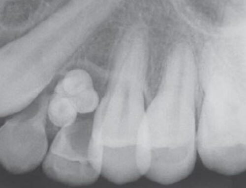
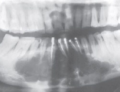
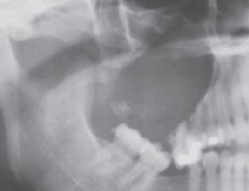
- Margins sharply defined, but can also be ill-defined.
- Often shows scalloping between roots.
Histopathology
- Never has an epithelial lining. Walls are lined by a thin band of vascular fibrous connective tissue or thickened myxofibromatous proliferation with reactive bone
Treatment
- Surgical exploration is needed to establish diagnosis. Diagnosis is primarily based on the clinical and radiographic features, together with surgical findings.
- Little or no tissue is obtained.
- Curettage of the bony walls.
- Recurrence or persistence of lesion is unusual.


