AKA
- Gorlin Cyst, Dentinogenic Ghost Cell Tumor, Calcifying Cystic Odontogenic Tumor, Calcifying Ghost Cell Odontogenic Cyst
Incidence
- mean age of 33; most cases diagnoses in 3rd and 4th decades of life
Clinical presentation
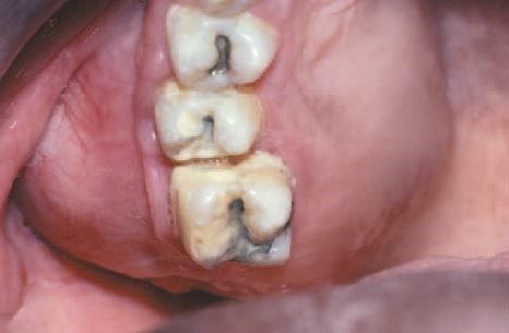
- extraosseous examples appear as localized sessile or pedunculated gingival masses with no distinctive clinical features.
Location
- 65% found in incisors and canines. Mandible = maxilla. Predominantly an intraosseous lesion, but 5% to 17% are extraosseous (peripheral).
Radiographic Features
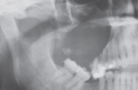
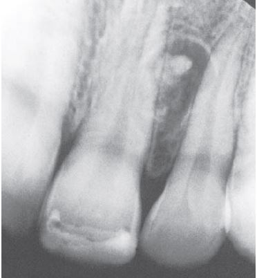
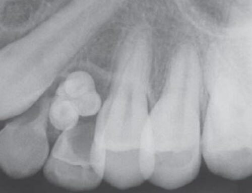
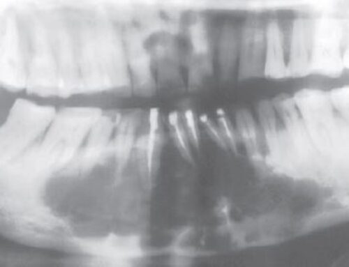
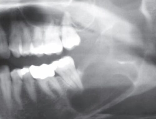
- Usually unilocular, well-defined radiolucency, although the lesion may occasionally appear multilocular. Radiopaque structures within the lesion, either irregular calcifications or toothlike densities, are present in about one third to one half of cases. Approximately one third of cases are associated with an unerupted tooth, most often a canine. Root resorption or divergence of adjacent teeth is sometimes seen.
Compare to
- Can be associated with odontomas, adenomatoid odontogenic tumors, ameloblastomas.
Histopathology
- well-defined cystic lesion with a fibrous capsule and a lining of odontogenic epithelium of 4-10 cells in thickness; basal cells of the epithelial lining may be cuboidal or columnar and are similar to ameloblasts; overlying layer of loosely arranged epithelium may resemble stellate reticulum of an ameloblastoma; eosinophilic “ghost cells” are characteristic – altered epithelial cells with loss of nuclei and preservation of the basic cell outline. Dystrophic calcifications may be present.
Treatment
- Prognosis good and recurrence low with simple enucleation.
Dentinogenic ghost cell tumors/epithelial odontogenic ghost cell tumors: have no cyst features, may be infiltrative or even malignant and are regarded as neoplasms
Calcifying odontogenic cysts (86-98%): represents nonneoplastic cysts

