AKA
- Pinborg Tumor
Incidence
- Accounts for less than 1% of odontogenic tumors. Most common from 30 to 50 years of age. No sex predilection.
Location
- 2/3 of cases in mandible, most often in posterior area
Origin
- The tumor bears close morphologic resemblance to the cells of the stratum intermedium of the enamel organ. Others suggest that the tumor arises from dental lamina remnants.
Clinical Features
- Painless, slow growing swelling is most common presenting sign. Peripheral (extraosseous) calcifying epithelial odontogenic tumors are sessile gingival masses on the anterior gingiva and have cupped-out erosions of the underlying bone.
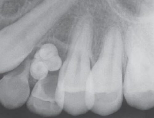
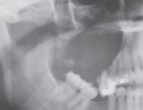
Radiographic Features
- Well-defined unilocular or multilocular radiolucent lesion, often scalloping and containing calcified structures. Tumor often associated with an impacted tooth.
Histopathology
- calcifications develop within the amyloid-like material and form concentric rings (Liesegang ring calcifications). Stains as amyloid and after Congo red staining, shows apple-green birefringence when viewed under polarized light
Treatment: conservative local resection to include a narrow rim of surrounding bone. Recurrence rate is 15%.


