Rule of 2/3rds: 2/3 occur in maxilla, 2/3 in second decade of life; 2/3 female
Cause
- Derived from enamel organ epithelium or from remnants of dental lamina.
Incidence
- represents 3% to 7% of all odontogenic tumors. 2/3 of cases occur in 2nd decade; female affected twice as often as males
Location
- often affects anterior jaw; maxilla affected twice as often as mandible
Radiographic Features
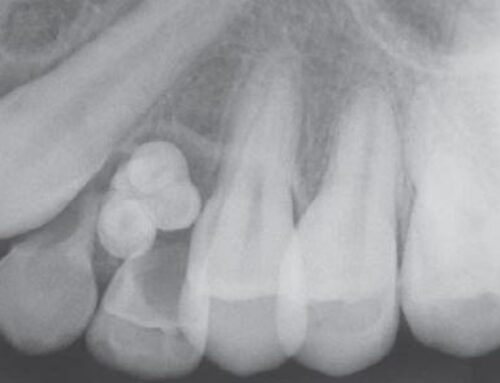
- in 75% of cases, circumscribed radiolucency that involves crown of unerupted tooth, most often a canine. Often contain fine (snowflake) calcifications. Follicular Type, Extrafollicular Type
Compare to
- Dentigerous Cyst
Histopathology
- well-defined lesion that is usually surrounded by a thick, fibrous capsule; may be solid or may show cystic changes; composed of spindle-shaped epithelial cells that form sheets, strands, or whorled masses of cells in a scant fibrous stroma; tubular or ductlike structures are characteristic and result from secretory activity of the tumor cells, which appear to be preameloblasts; small foci calcifications may be scattered through the tumor.
Treatment
- capsules allows for easy enucleation
Follicular Type of Adenomatoid Odontogenic Tumor
- Involves crown of unerupted tooth. Sometimes extends apically along the root past the CEJ, which helps distinguish it from a dentigerous cyst (Fig 15-85)
Extrafollicular Type of Adenomatoid Odontogenic Tumor
- Well-delineated unilocular radiolucency that is not related to an unerupted tooth (Fig 15-86)


